* These statements have not been evaluated by the Food and Drug Administration. This product is not intended to diagnose, treat, cure, or prevent any disease. For research purposes only.
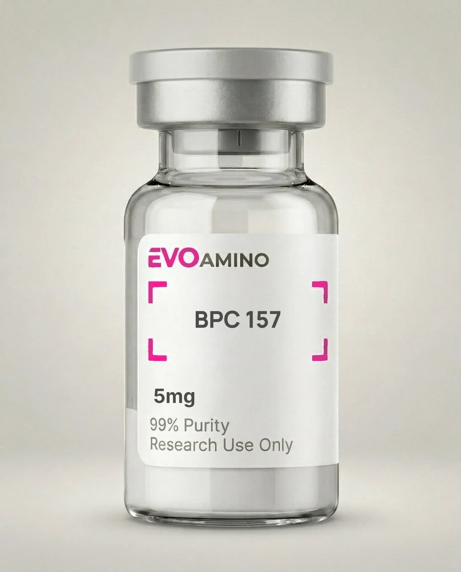
The molecular architecture of BPC-157 begins with a 15-residue linear sequence: Gly-Glu-Pro-Pro-Pro-Gly-Lys-Pro-Ala-Asp-Asp-Ala-Gly-Leu-Val. Published structural analyses confirm the molecular formula as C₆₂H₉₈N₁₆O₂₂ with a molecular weight of 1419.53 g/mol and CAS number 137525-51-0 (PMID: 30915550). The peptide derives from a partial sequence of human gastric juice protein, specifically a protective protein fraction within gastric mucosa. Three consecutive proline residues at positions 3–5, plus a fourth at position 8, introduce marked conformational constraints that restrict rotational freedom along the backbone. N-terminal glycine and C-terminal valine define the bookend residues. Published NMR spectroscopy studies reveal that BPC-157 adopts a partially helical conformation in aqueous solution, with more ordered structure at the central region and flexible termini. Solid-phase Fmoc chemistry is the synthesis route of choice, followed by preparative HPLC purification and lyophilization. The resulting lyophilized powder is white to off-white and freely soluble in water and aqueous buffers — properties that support its broad utility as a preclinical research tool.
What is the origin of BPC-157?
BPC-157's origin lies in the isolation of a protective protein fraction from human gastric juice. Investigators searching for endogenous cytoprotective factors in gastric mucosa identified this protein fraction based on its activity in tissue protection models. The 15-amino acid fragment was subsequently characterized as the minimal active sequence accounting for the observed protective effects. The designation "Body Protection Compound" encodes the original discovery context — a molecule native to stomach tissue that guarded against mucosal injury. Published studies place this discovery in the early 1990s, with characterization work establishing isolation criteria and protective activity benchmarks in gastric tissue models. What distinguishes BPC-157 from most research peptides is that it is not a de novo synthetic construct — it mirrors a naturally occurring sequence in human tissue. Published molecular biology studies confirm that the synthetic version is physicochemically equivalent to the native fragment. That endogenous origin gives BPC-157 a particular relevance in gastric biology research contexts and positions it as a model compound for studying cytoprotection at the molecular level.
What are the published mechanisms of action for BPC-157?
BPC-157 engages multiple interacting molecular pathways, with the nitric oxide (NO) system representing the most extensively characterized primary target. Studies in endothelial cell cultures and gastric tissue models document effects on NO synthesis and downstream signaling. Endothelial nitric oxide synthase (eNOS) activity is modulated, with downstream effects on vascular tone and blood flow regulation in these in vitro systems. The GABAergic system represents a second documented pathway: published in vitro studies show effects on GABA-A receptor function and GABAergic neurotransmission in neuronal cell cultures. Dopaminergic pathway modulation has been characterized in cell culture models, with effects on dopamine synthesis, transporter function, and receptor activity. Growth factor signaling — particularly VEGF and FGF — is affected in tissue culture studies, with documented changes in cell proliferation and migration responses (PMID: 30915550). BPC-157 also intersects the prostaglandin system, modulating prostaglandin E2 synthesis in epithelial cell cultures. Published literature additionally describes activity at the NO-cGMP axis and calcium signaling components, underscoring the mechanistic breadth that makes BPC-157 a useful tool for multi-pathway research designs.
What does published research show about BPC-157 and nitric oxide?
The nitric oxide system emerges as a primary molecular interface for BPC-157 across published in vitro work. Endothelial cell culture studies demonstrate that BPC-157 affects eNOS expression and NO output, with downstream consequences for vascular tone and cellular communication. NO is a short-lived but potent signaling molecule governing smooth muscle relaxation, platelet aggregation, and endothelial homeostasis. Published molecular studies in gastric tissue models show that BPC-157 influences NO-mediated protective responses against injury, with the peptide appearing to enhance NO bioavailability through effects on synthesis and tissue-level NO stabilization. The NO-cGMP pathway represents an additional documented axis: published research characterizes effects on cyclic GMP production downstream of NO signaling, with secondary consequences for smooth muscle relaxation and cellular stress responses. Tissue culture studies confirm that BPC-157's influence on NO signaling correlates with measurable changes in cellular functional outputs. Published mechanistic reviews treat NO pathway modulation as the principal molecular mechanism through which BPC-157 exerts its documented effects in preclinical models — a conclusion supported by the volume and consistency of in vitro evidence across multiple laboratory systems.
How does BPC-157 affect growth factor pathways?
Growth factor signaling represents a mechanistically distinct dimension of BPC-157 research. Published studies in fibroblast and endothelial cell cultures document effects on VEGF expression and VEGF receptor signaling, with measured changes in angiogenic responses in vitro. VEGF-A production appears elevated in cellular models following BPC-157 exposure, with potential downstream consequences for vascular formation and remodeling processes. FGF-2 signaling is a second documented target: published work describes effects on fibroblast proliferation and extracellular matrix production in cell culture systems, with effects mediated through FGF receptor activation and downstream MAP kinase signaling. EGF receptor phosphorylation is also affected, with published scratch assay data in epithelial cell cultures showing changes in cell migration and wound closure kinetics following BPC-157 treatment. Published molecular summaries characterize this growth factor signaling cross-talk as a mechanistic route through which BPC-157 influences cell proliferation, migration, and tissue remodeling responses in preclinical systems (PMID: 30915550). These pathways — particularly their overlap with angiogenesis and tissue repair cascades — are the primary reason BPC-157 appears in connective tissue and vascular biology research designs.
What is known about BPC-157 and neurotransmitter systems?
BPC-157 has been characterized across three major neurotransmitter systems in published in vitro research: GABA, dopamine, and serotonin. Neuronal cell culture studies document that BPC-157 affects GABA-A receptor function, with measurable effects on chloride channel conductance and GABAergic signal transduction. Receptor subunit expression and trafficking are both affected in cellular models. Dopaminergic pathways represent a second characterized system: published work shows BPC-157 influences dopamine synthesis, release, receptor activity, and transporter function in neuronal cultures. Serotonin system interactions are documented but less fully characterized than the GABA and dopamine data. Published molecular research suggests that neurotransmitter metabolism is altered through effects on synthetic enzymes — monoamine oxidase activity in biochemical assays shows sensitivity to BPC-157, pointing to potential effects on neurotransmitter catabolism rates. Published studies are consistent in framing these findings as mechanistic data from cell culture and biochemical models, providing pathway-level insights that represent preliminary evidence requiring independent replication before more definitive mechanistic conclusions can be drawn.
What is the amino acid sequence of BPC-157?
The primary structure of BPC-157 is Gly-Glu-Pro-Pro-Pro-Gly-Lys-Pro-Ala-Asp-Asp-Ala-Gly-Leu-Val, a 15-residue linear arrangement. Single-letter notation: GEPPPGKPADDAGLV. Published structural studies confirm this sequence through both mass spectrometry and Edman degradation sequencing. The sequence encodes several structurally and chemically significant features. The Pro-Pro-Pro triplet at positions 3–5 introduces polyproline-type conformational rigidity, a structural property that distinguishes BPC-157 from most short research peptides. Multiple acidic residues — glutamic acid at position 2 and aspartic acid at positions 10–11 — govern charge and solubility behavior. Hydrophobic residues leucine and valine at the C-terminus, alongside alanine, balance the hydrophilic character of the charged residues. Glycine at the N-terminus permits backbone flexibility. No cysteine residues are present, eliminating disulfide bond formation and simplifying both synthesis and long-term stability management. Published solid-phase synthesis studies note that the proline-rich segment requires careful coupling optimization to prevent incomplete reaction. Circular dichroism spectroscopy data indicate random coil and partial helical structure in solution, consistent with NMR observations of the central helical region.
How does BPC-157 interact with cellular stress responses?
Cellular stress response pathways — oxidative stress, ER stress, and mitochondrial function — each show documented sensitivity to BPC-157 in published in vitro work. Epithelial cell culture studies demonstrate effects on antioxidant enzyme expression: superoxide dismutase and catalase levels are affected, with downstream changes in cellular redox status. Reactive oxygen species production and scavenging dynamics are both modulated in oxidative stress model systems. Heat shock protein expression is a second documented endpoint: HSP70 and HSP90 levels shift in cellular stress models following BPC-157 treatment, affecting the molecular chaperone network that supports protein folding under stress conditions. Published work also characterizes effects on the unfolded protein response, with ER stress markers showing sensitivity to BPC-157 in cell culture systems. Mitochondrial studies add a third mechanistic layer: effects on membrane potential and ATP production are documented, along with influence on mitochondrial biogenesis markers and mitochondrial dynamics. These parallel findings across multiple cellular stress pathways provide mechanistic context for the cytoprotective properties repeatedly observed in tissue culture studies and inform experimental designs targeting multi-stress endpoints.
What research applications does BPC-157 have?
The research application space for BPC-157 spans several in vitro domains, each driven by the mechanistic profile detailed in published literature. Gastric mucosal biology research uses BPC-157 to probe protective mechanisms against chemical and oxidative injury in mucosal cell cultures. Wound healing applications leverage its effects on fibroblast migration, collagen synthesis, and angiogenic signaling. Vascular biology research examines endothelial cell function, angiogenesis, and vascular remodeling in vitro. Neuroscience applications use BPC-157 to interrogate neurotransmitter modulation and neuronal stress responses in cell culture formats. Connective tissue research — tendons and ligaments specifically — investigates BPC-157 effects on connective tissue cell behavior and extracellular matrix production. Published studies consistently frame these applications as preclinical research using cell cultures, tissue explants, and biochemical assays rather than controlled clinical investigations. Experimental protocols typically use 1–100 μg/mL in cell culture media, with exact concentrations selected based on cell type, endpoint, and published precedent. High-purity compounds with verified sequences, HPLC purity documentation, and mass spectrometry identity confirmation are standard requirements for reliable data generation in these applications.
What is the current state of BPC-157 research?
The published BPC-157 literature base is predominantly preclinical, comprising cell cultures, tissue models, and biochemical assays. Published review articles characterize this literature as mechanistically informative but not yet providing the rigorous clinical validation required for definitive conclusions. NO pathway modulation, growth factor signaling, and neurotransmitter system interactions represent the best-characterized mechanistic territory. Published findings span gastric, vascular, and neuronal tissue culture systems. The notable gap in the literature is the absence of large-scale randomized controlled investigations, which limits the interpretive weight of existing mechanistic findings. Published research calls for additional work to clarify molecular targets and define signaling hierarchy across the multiple pathways BPC-157 engages. Methodological limitations and replication gaps are noted in published quality assessments of the field. Systematic reviews flag the need for standardized experimental protocols and independent replication of key mechanistic findings before stronger conclusions are warranted. Research-grade BPC-157 from documented-quality sources enables investigators to conduct mechanistic studies using well-characterized compounds — a prerequisite for producing data that can meaningfully advance the field.
FAQ
What is the molecular weight of BPC-157?
The molecular weight of BPC-157 is 1419.53 g/mol (monoisotopic) or 1419.64 g/mol (average). Published mass spectrometry studies confirm this molecular weight with high accuracy.
What is the CAS number for BPC-157?
The CAS Registry Number for BPC-157 is 137525-51-0. This unique identifier distinguishes it from related compounds and provides standardized reference for chemical databases and regulatory documentation.
How stable is BPC-157 during storage?
Lyophilized BPC-157 is stable at -20°C for 24+ months. The peptide is susceptible to oxidation and hydrolysis in solution. Published stability studies recommend aliquoting into single-use volumes and storing at -20°C or -80°C (PMID: 30915550).
What concentration is used in cell culture research?
Published in vitro studies typically use BPC-157 concentrations of 0.1-100 μg/mL, with 1-10 μg/mL being most common. Concentrations vary by cell type and experimental design. Always verify viability at planned concentrations.
Does BPC-157 form disulfide bonds?
No, BPC-157 contains no cysteine residues and cannot form disulfide bonds. This simplifies synthesis and handling compared to disulfide-containing peptides. The linear structure remains the active form.
Research Use Only: All compounds sold by Evo Amino are intended exclusively for laboratory research. Not for human or animal consumption. These products are not drugs, supplements, or food. Statements have not been evaluated by the FDA. Must be 21+ to purchase.
Cited literature
References
Primary literature and public databases referenced above. Each link resolves on the publisher or database of record.